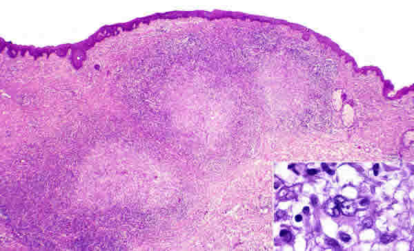
Fig. 1: Dermal infiltrate showing nodular aggregates of tumour cells bordering a central area of necrosis. (Haematoxylin-eosin stain x40). Inset shows a high power of the neoplastic cells with abundant cytoplasm, irregular nuclei and prominent nucleoli. (Haematoxylin-eosin stain x400).