
Vol. 42, n.º 2, 2009
|
REVISTA
ESPAÑOLA DE
Vol. 42, n.º 2, 2009 |
COMUNICACIONES BREVES
Ricardo Drut, Eugenia Altamirano, Alejandra María Ollano
Department of Pathology. «Superiora Sor María
Ludovica» Children’s Hospital. 1900 La Plata. Argentina
patologi@netverk.com.ar
SUMMARY
For the first time, to our knowledge, we report a peculiar histological finding in the thyroid glands of children, namely the presence of empty, elongated, tortuous, retraction-like spaces apparently dissecting the interstitial tissue. The availability of D2-40, an immunohistochemical marker for lymphatic endothelial cells, allowed us to identify these spaces as intraparenchymatous lymphatic capillaries. This phenomenon was evident at the periphery where they blended with marginal lymphatic vessels.
Keywords: D2-40, lymphatic capillaries, thyroid gland.
RESUMEN
En esta comunicación presentamos el peculiar hallazgo histológico de cavidades vacías, alargadas, tortuosas, de aspecto artefactual y aparentemente disecando el intersticio en tiroides de niños. La disponibilidad del marcador inmunohistoquímico D2-40 para células endoteliales de linfáticos permitió reconocer que los referidos espacios representan capilares linfáticos intraparenquimatosos. Este fenómeno fue más obvio en la periferia de la glándula donde estas cavidades se unían con los linfáticos marginales. No hemos podido encontrar referencia alguna mencionando estos hallazgos.
Palabras clave: Capilares linfáticos, D2-40, tiroides.
INTRODUCTION
In recent years we have noticed a peculiar histological pattern in the thyroid glands of autopsy specimens of newborn infants. The cracking-like pattern was not uniformly distributed, but was always found to be present when specifically looked for. Although at the periphery of the gland the spaces seemed to belong to endothelial-lined vascular structures, most probably lymphatic capillaries, the spaces within the gland followed extremely irregular and tortuous outlines, focally dissecting local structures. The resulting images strongly suggested retraction artefacts. The aim of the present study was to identify the origin of these spaces.
MATERIALS AND METHODS
Paraffin blocks from autopsy samples of transversally sectioned trachea with the thyroid gland lobes on both sides were retrieved from the files of the department of pathology, comprising of 7 newborns (up to 28 days), 7 infants (1-12 months), and 7 children (13 months to 15 years).
The histology of the blocks was carefully re-evaluated and new sections immunostained for D2-40 (DakoCytomation, Code M3619, dilution 1/100, with citra solution antigen retrieval, 2 x 20 minutes) as a marker for lymphatic endothelial cells (1-3).
RESULTS
All the thyroid glands presented different degrees of the changes described in the introduction. The empty spaces were elongated and followed a somewhat sinuous course; most appeared to be devoid of a cellular lining. Some spaces were arranged around groups of follicles of different size. In some cases the cavities were so marked that the groups of follicles resembled pieces of a jigsaw puzzle. A dissecting pattern around small pieces of collagen-containing interstitial connective tissue was also seen (fig. 1). At the periphery of the gland the empty spaces were continuous with periglandular lymphatic capillaries with an obvious endothelial lining (fig. 2).
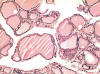
Fig. 1:
The peculiar empty interstitial spaces as seen in different thyroid glands.
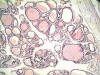
Fig. 2:
Empty spaces joining a peripheral lymphatic vessel.
D2-40 immunostaining produced a brisk reactivity in the form of a line at the luminal side of the cavities and also stained the endothelial cells of the peripheral lymphatic capillaries (figs. 3 and 4).
![]()
Figs. 3 and 4:
The lining in the apparently empty spaces is D2-40 positive.
DISCUSSION
Although the occurrence of intrathyroid lymphatic capillaries is well described in the literature (4), no particular reference is made to the histological pattern of these vessels. The peculiar, almost jigsaw puzzle-like, pattern found in some cases seems to indicate that lymphatic capillaries are indeed numerous within the parenchyma of the gland. Although extremely difficult to detect by routine H&E staining, immunostaining with D2-40, a well-known marker for lymphatic endothelial cells, highlights the endothelial lining of the lymphatic vessels. However, no evidence of the pattern was found in adjacent tissues or organs such as the parathyroid gland.
In conclusion, the thyroid gland of children contains a peculiar and prominent complex lymphatic network which frequently appears extremely dilated. These lymph vessels can be recognized when stained with D2-40.
Although some spaces resembling the present findings can be seen in the illustrations of thyroid glands of fetuses and newborns in the book by Valdés-Dapena, no description of them is made in the text (5).
The abundance of intraparenchymatous lymph vessels in the thyroid gland is related, most probably, to the transport of the local hormones, an established phenomenon. It may help to explain the early metastatic involvement of local lymph nodes in the presence of thyroid carcinoma.
REFERENCES
Ordoñez, NG. Podoplanin: a novel diagnostic immunohistochemical marker. Adv Anat Pathol 2006; 13: 83-8.
Fukunaga M. Expression of D2-40 in lymphatic endothelium of normal tissues and in vascular tumours. Histopathology 2005; 46: 396-402.
Galambos C, Nodit L. Identification of lymphatic endothelium in pediatric vascular tumors and malformations. Pediatr Dev Pathol 2005; 8: 181-9.
Carcangiu ML, Thyroid. In Sternberg S. (ed.) Histology for pathologists. 2nd Edition. Philadelphia: Lippincott-Raven; 1997, p. 1077.
Valdés-Dapena MA. Histology of the fetus and newborn. Philadelphia: W.B.Saunders; 1979, p. 117.
![]()